Imaging of the molecular distributions at the cellular level (IMAGOCELL)
Type of project: national project
Duration: 2019 - 2022
Project leader: Prof. Dr Primož PeliconCode: ARRS N1-0090Coworkers: Doc. Dr Klemen Bučar, Prof. Dr Katarina Vogel-Mikuš, Doc. Dr Paula Pongrac, Dr Primož Vavpetič, Dr Boštjan JenčičPartners: Jožef Stefan InstituteInfrastructure: MIC - Microanalytical Centre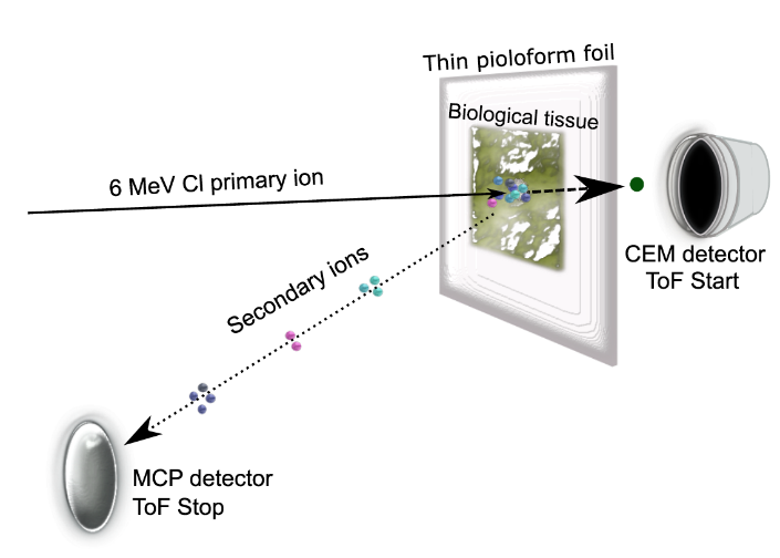
In a search for Imaging Mass Spectrometry (IMS) technique, which would be able to probe tissue with subcellular resolution, the detection of secondary molecular ions with mass spectrometer needs to be coupled with a soft probing mechanism, allowing the survival of large organic molecules during desorption from the sample, and efficient ionization of the desorbed molecules to enable their detection in mass spectrometers. The technology that best fulfils these criteria is based on secondary ion emission induced by impact of swift heavy ions on insulating material: heavy ions with energy in the MeV range (swift ions) interact with the matter by exchanging energy almost exclusively with electrons. The cylindrically shaped damaged region along the straight path of swift ion in the insulator featuring the burst of high electron temperature, has a diameter typically from one to four nanometres. Outside this region, a phonon shock wave propagates through the surrounding material, induces specific soft desorption process from the near-surface volume (referred to either as electron desorption, or electron sputtering), and surprisingly results in a high fraction of ionized non-fragmented large organic molecules. The IMAGOCELL project explores the physics behind the MeV-SIMS process to develop the methodology for advanced applications in biology and medicine.