Molecular imaging inside the cell (MICE)
Type of project: national project
Duration: 2018 - 2021
Project leader: Prof. Dr Primož PeliconCode: ARRS J7-9398Coworkers: Doc. Dr Klemen Bučar, Prof. Dr Katarina Vogel-Mikuš, Dr Mitja Kelemen, Doc. Dr Paula Pongrac, Dr Boštjan JenčičExternal coworkers: Dr Marjana Regvar, BF UL Dr Mateja Germ, BF UL Dr Matevž Likar, BF UL Mateja Potisek, young researcher, BF UL Anja Kavčič, young researcher, BF ULInfrastructure: MIC - Microanalytical Centre
Imaging Mass Spectrometry (IMS) provides insight into biomolecular processes by imaging of molecular distributions inside the biological tissue, revealing the complex physiological processes inside living organisms. Currently available IMS technologies lack of abilities to image the molecular distributions at subcellular level, as the physical processes of molecular desorption either destroy the fragile chemical bonds and defragment the molecules, or are not capable to desorb the molecules from limited area of the sample. The project MICE (Molecular Imaging Inside The Cell) is engaged in developing a new IMS technology, which incorporates the efforts and knowledge from the field of physics, biomedicine and engineering. As a basic physical process behind the thechnology we exploit the electron desorption phenomenon, induced by heavy, high energy ion impact on the surface of the biological tissue.
The superior IMS technology is known as MeV-SIMS (Secondary Ion Mass Spectrometry with MeV ions).
The project consists of four working packages, each encompassing the following tasks:
- WP1: From 10-μm to 500-nm resolution IMS technology (T1: Tissue processing for subcellular IMS with continuous beam, T2: Forming of 500-nm swift ion beam, T3: New sequence of TOF imaging: start by primary ion arrivals)
- WP2: Imaging of intracellular biochemistry (T1: Imaging the paths of gene expression in plant cells, T2: Response in cells to synthetic compounds and drug applications.
- WP3: Improving the mass spectrometry (T1: Upgrading the TOF mass spectrometer: from linear TOF to reflectron. T2: Upgrade of FPGA electronics: from 4 ns down to 300 ps timing resolution)
- WP4: Outreach of technology, exploitation, dissemination (T1: Pathways of access to the new IMS technology, T2: Industrial applications, dissemination, exploitation)
After the Month 23 of the ongoing project (as of May 1, 2020), the following progress is achieved within the listed Work Packages:
WP1:
T1: The tissue processing technology for conventional MeV-SIMS (similar to standard SIMS technique) has been completely changed. In order to be able to detect swift ions behind the sample, we applied thin cuts at the cryotome (i.e. 10 micrometer thick slice of vitrified frozen hydrated tissue), deposit it on ultra-thin conductive membrane (100 nanometer coated polymer foil), and freeze-dry it. In this way, after the removal of water, the resulting thickness of dry tissue of app. 4 micrometres allows for the penetration of the 6 MeV Cl ions. The technique was applied and confirmed to function both at the human liver tissue, as well as on corn seed tissue.
T2: The formation of the heavy high energy ion beams is at Ljubljana tandetron associated with the use of sputter ion source with limited beam brightness, which determines the quality of the focused beam. In order to be able to operate with the beam with sub-micrometer 35Cl6+ (clorine ion beam with ion charge 5+), energy of cca 5 MeV or higher, the beam diameter is reduced by closing the object and collimator slits at the microprobe. As a consequence, the beam intensity dropped for five orders of magnitude, from 109 ions/sec to 104 ions/sec. The beam diameter was measured with mesh scanning and channeltron positioned, and optical parameters tuned. Beam diameter of 800 nanometers was achieved at the ion fluxes of approx. 5000 ions/sec.
T3: At such low beam intensities, each ion should be used to desorb the ions for the mass spectrometry to achieve considerable yield in the mass spectra. In the standard mode of operation, the continuous beam is changed into pulsed by fast deflectors installed in the beamline. This would not work with such low intensity beam. New mode of Time-Of-Flight (TOF) operation is successfully applied within MICE project. Continuous electron multiplier (channeltron) is installed behind the sample, detecting each arrival of the primary ion beam. TOF measurements is triggered (TOF start) at each arrival, and secondary ion travel time within the linear TOF spectrometer (TOF stop) measured. The “TOF start” definition is in this case excellent, with the precision within 1 nanosecond, resulting in improved mass resolution.
WP2:
T1: Work in plant biology with MeV-SIMS is ongoing in parallel with the development of the method. The existing state-of-the-art MeV-SIMS technology is explored to map secondary metabolite distributions in plant tissue. One of the objects extensively investigated is buckwheat plant, locally important staple food, and a novel “superfood” candidate due to presence of strong antioxidants. We focused on the molecular mapping of rutin and quercetin in buckwheat/tartary buckwheat seeds, providing insight into mill processing potential of buckwheat seed fractions.
T2: In order to understand the penetration of pesticides into food products, interaction of corn with known herbicide glyphosate was investigated. Distribution of the glyphosate in the corn seed tissue was measured with lateral resolution of 800 nanometers. First indication drawn were, that the outer layer contains higher concentrations of the glyphosate.
WP3:
T1: Extensive design study was carried out in order to extend the flight path of the secondary ions within the TOF mass spectrometer (TOF MS), and to add additional reflectron stage. Public tender for the upgrade of the TOF MS was executed in the end of 2019, and the reflectron stage arrived in the beginning of 2020. Installation of the reflectron stage is ongoing at the moment.
T2: Complete renewal of the acquisition system at the high energy focused ion beam at JSI took place in the send half of 2019 and in the beginning of 2020. Installation of the new hardware and software was executed by engineer of the selected company. Integration of the ultra-fast TDC with timing resolution of 250 picosecond was done during the campaign, and the operation tested/verified in the beginning of February 2020.
WP3, extended activities:
As the detection efficiency of microchannel-plate detector for the heavy secondary ions with masses exceeding 1000 Da and travel energies of 3 keV is very low, alternative post-acceleration approaches and detection approaches were studied. A dedicated discrete anode detector for secondary ions with 10 keV post-acceleration is selected, and acquired. We are now anticipating efficient detection of the secondary ions with masses exceeding 3 kDa at extraction energies of 3 keV.
WP4:
T1: MeV-SIMS in its existing state-of-the-art is including in the Transnational access programme funded by the EU within the H2020 project “Radiate”, which is merging the capabilities of 15 top European ion beam facilities. In addition, it is a part of the techniques contributed to the consortium SiMBion, a national node of Euro-BioImaging ESFRI (ERIC) project. Both EU frameworks offer (will offer) the access of the European researchers to the analytical capabilities of the MeV-SIMS.
T2: Communication with potential industrial customers is taking place. Penetration of the pesticides into fruits is one of the topics addressed in the next months.
Project references:
WP1, WP2: Boštjan Jenčič, Primož Vavpetič, Mitja Kelemen, Matjaž Vencelj, Katarina Vogel-Mikuš, Anja Kavčič, Primož Pelicon, MeV-SIMS TOF Imaging of Organic Tissue with Continuous Primary Beam, Journal of the American Society for Mass Spectrometry 30 (2019) 1801-1812.
https://doi.org/10.1007/s13361-019-02258-8
WP1, WP3: Boštjan Jenčič, Primož Vavpetič, Mitja Kelemen, Primož Pelicon, Secondary Ion Yield and Fragmentation of Biological Molecules by Employing 35Cl Primary Ions within the MeV Energy Domain, Journal of the American Society for Mass Spectrometry 31 (2020) 117-123.
https://doi.org/10.1021/jasms.9b00019
WP4:
S. Hoereth et al, Arabidopsis halleri shows hyperbioindicator behaviour for Pb and leaf Pb accumulation spatially separated from Zn, New Phytologist 226 (2020) 492–506.
https://doi.org/10.1111/nph.16373
P. Pongrac et al, Contrasting allocation of magnesium, calcium and manganese in leaves of tea (Camellia sinensis (L.) Kuntze) plants may explain their different extraction efficiency into tea, Food and Chemical Toxicology 135 (2020) 110974.
https://doi.org/10.1016/j.fct.2019.110974
Malay et al, An ultra-stable gold-coordinated protein cage displaying reversible assembly, Nature 569 (2019) 439.
https://doi.org/10.1038/s41586-019-1185-4
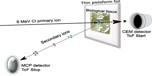
Figure 1: MeV-SIMS with continuous beam (Source: Jenčič et al, JASMS 2019).
Figure 2: MeV-SIMS molecular fragmentation trend: fragmentation probability of large organic molecules is falling with the increasing projectile energy (Source: Jenčič et al, JASMS 2020).