An important acquisition for the XRF laboratory
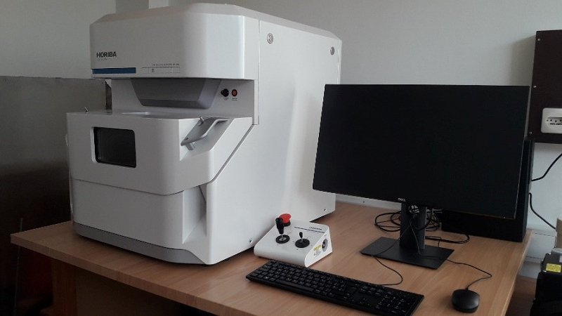
Micro-XRF (X-ray fluorescence) spectrometry is imaging technique which enables localization of elements in different solid samples: biological tissues, geological materials, metals, cultural heritage artefacts. Our XRF laboratory (Laboratory for X-ray fluorescence spectrometry) is now equipped with the state of the art micro-XRF analytical microscope XGT-9000, enabling the team to scan samples sized from few mm up to 15 cm. The first measurements were successfully performed:
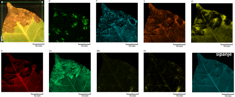
Figure above: Coffee (Coffea arabica L.) leaf, infected by pathogenic fungi. Upper left: visible-light stereomicroscope photograph. Lateral resolution ~140 um. Frame size 35 mm x 33 mm, 256 x 243 px, mapping time 58 min.
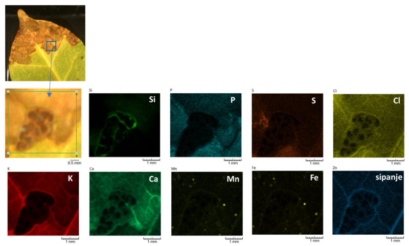
Figure above: Coffee (Coffea arabica L.) leaf, infected by pathogenic fungi – zoom of fungal sporocarp. Upper left: visible-light stereomicroscope photographs with marked zoom region. Lateral resolution ~15 um. Frame size 3,6 mm x 4 mm, 256 x 230 px, mapping time 43 min.