Circular trap mass spectrometer (orbitrap) and MeV-SIMS
Mass spectrometry made an important breakthrough with the invention of the circular trap mass spectrometer. With this discovery, the mass resolution exceeded the limit where different molecular ions with the same number of nucleons can be easily separated. In the seminar, we will examine how a circular trap spectrometer works. We will try to understand why it took 76 years of unsuccessful experiments from Kingdon's idea [1] in 1923 to make a working circular trap mass spectrometer, which Makarov succeeded in 1999. Can a circular trap spectrometer be used for imaging mass spectrometry? How to use a circular trap spectrometer to image the distribution of organic macromolecules in biological tissues using the MeV-SIMS method [3]? How to combine such a spectrometer with an ion microbeam on an IJS accelerator?
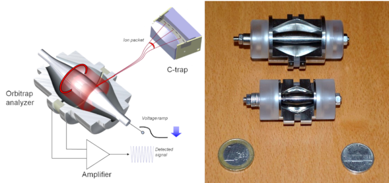
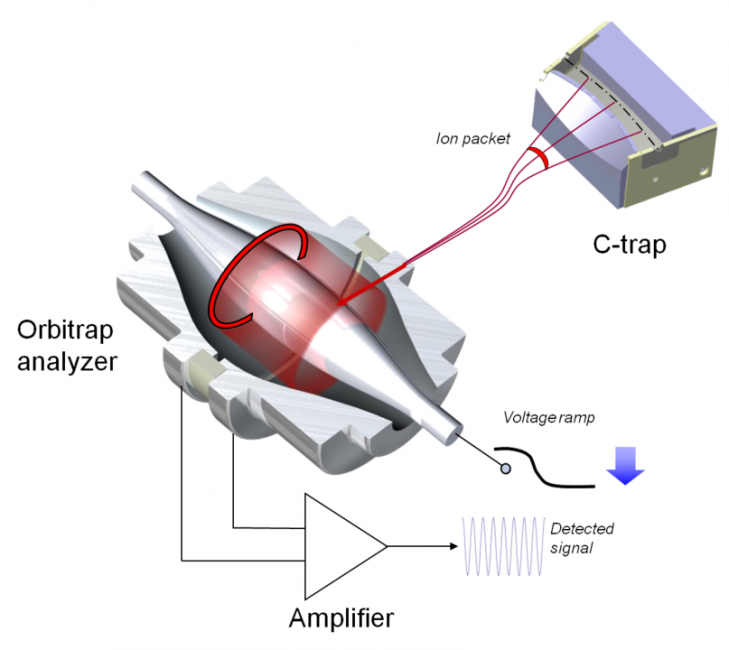
Figures above: schematic operation of a circular trap spectrometer and a photograph of the most important part of the spectrometer (source: Thermo Fisher Scientific (Bremen), CC BY-SA 3.0, https://en.wikipedia.org/wiki/Orbitrap#cite_note-Mak1-1).
[1] Kingdon KH (1923). "A Method for the Neutralization of Electron Space Charge by Positive Ionization at Very Low Gas Pressures". Physical Review. 21 (4): 408–418.
[2] Makarov, A (2000). "Electrostatic axially harmonic orbital trapping: A high-performance technique of mass analysis". Analytical Chemistry. 72 (6): 1156–62.
[3] Jenčič, B, et al (2019). “MeV-SIMS TOF imaging of organic tissue with continuous primary beam”. Journal of the American Society for Mass Spectrometry. 30 (9): 1801-1812.